Technique Development
Many of our research efforts focus on developing new methods for real-time studies using electron microscopy methods. The philosophical motivation for this is described in an article on how science advances by A.M. Schneider entitled “Four stages of a scientific discipline: four types of scientists“. He posits that there are four stages of research:
- Identification of the object of research
- Development of tools/instruments to investigate the underlying phenomena
- Investigation of the research by the developed tools, and
- The codification and refinement of knowledge.
Below, you will find short descriptions of some of our work concerning Stage 2. Stage 3 research work is described on the Research page.
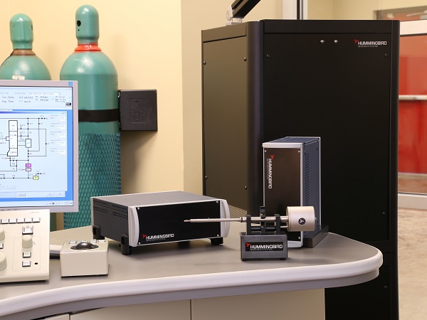
High-pressure catalysis
In atmospheric pressure microscopy, we use closed-cell methods similar to those for liquid microscopy to provide both high temperatures and pressures reaching up to 40 bar. This allows us to understand how the size, structure and composition of supported heterogeneous catalyst change in reactive environments.
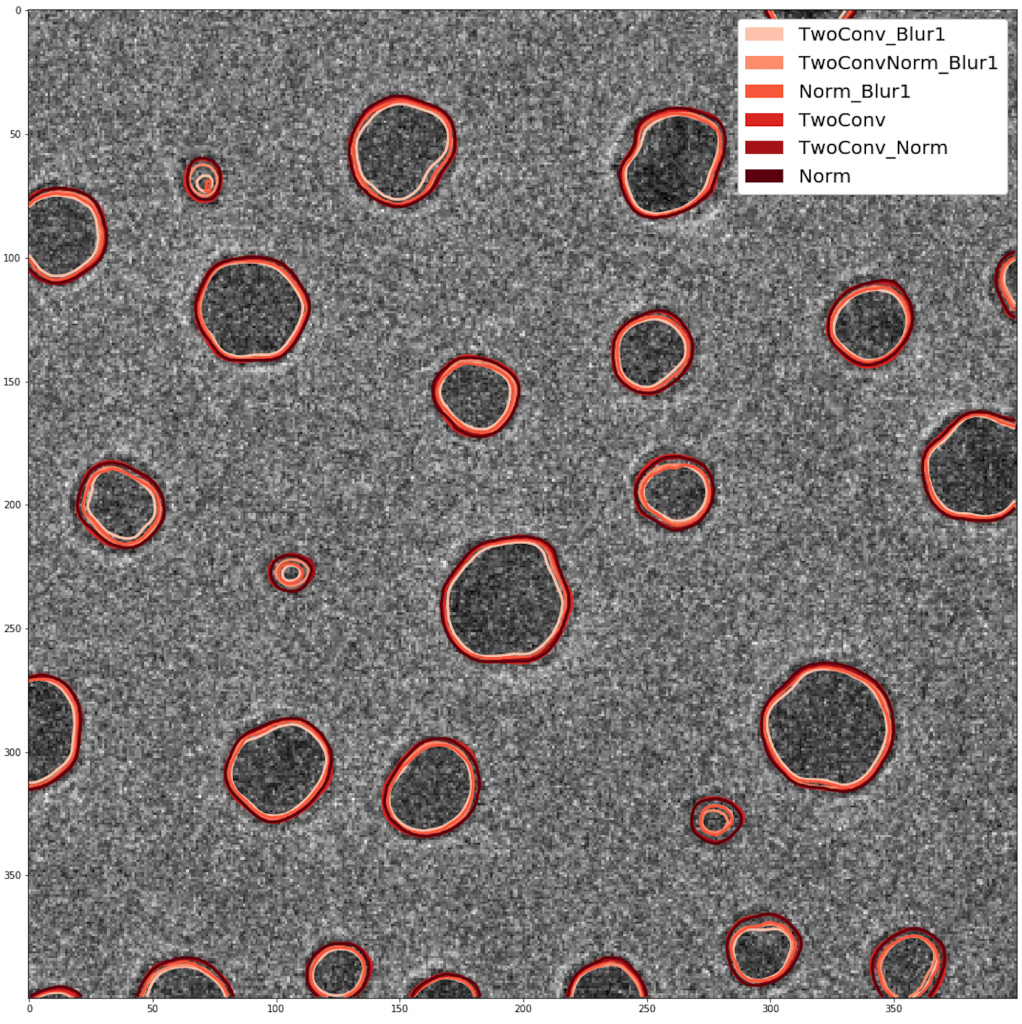
Machine Learning
We apply state-of-the-art machine learning techniques to problems in electron microscopy. Initial work focuses on developing deep-learning methods for binary image segmentation to aid the processing of electron micrographs. While our modern direct electron detection cameras enable imaging hundreds of frames per second, the amount of data we extract is limited by the number images that can actually be processed – our recent results have shown that applying a convolutional neural network to images of supported nanoparticles can reduce the time required to identify and measure particles from over 12 hours per image to merely 0.5 seconds per image.
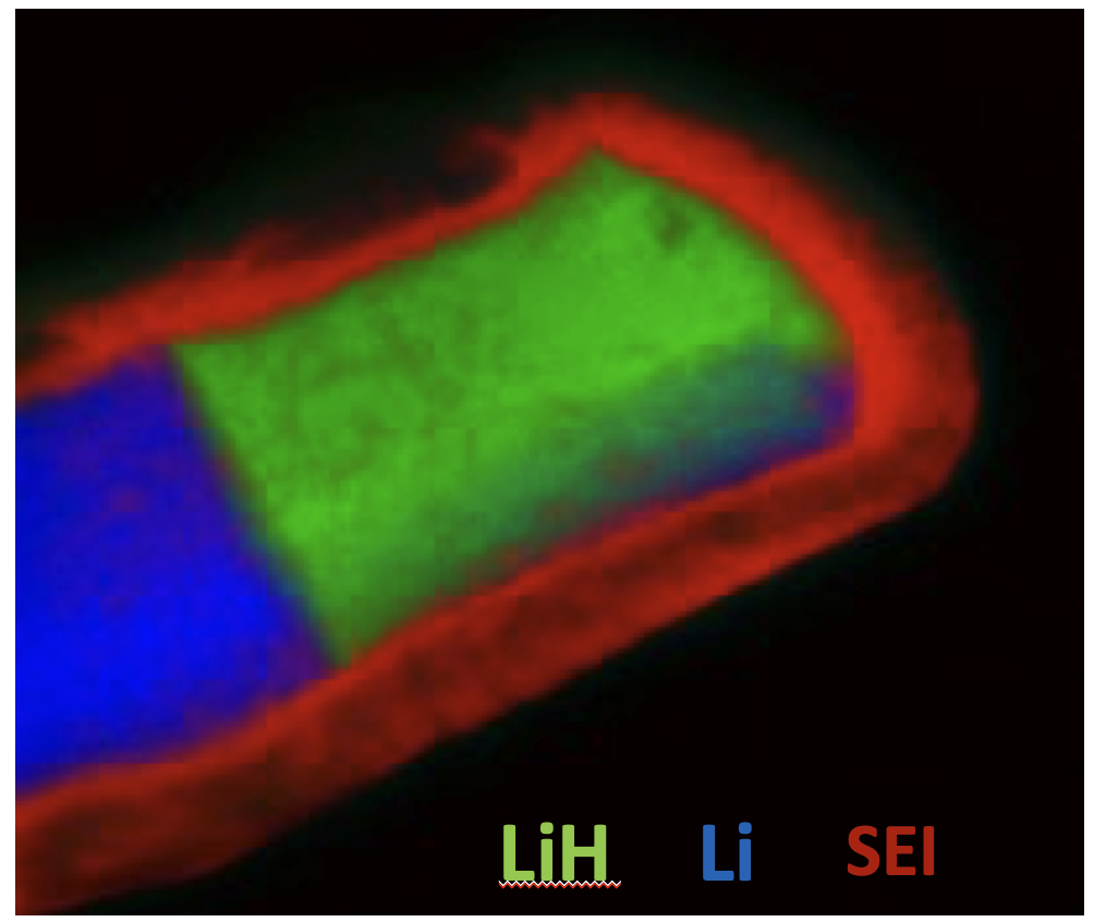
Cryogenic Electron and Ion Microscopy
Soft matter is very often damaged by intense electron irradiation in the transmission electron microscope. Our group has developed workflows for the characterization of soft matter using cryogenic ion and electron microscopy and low dose methods. Applications include characterization of the solid-electrolyte interphase in batteries as well as soft matter systems relevant to LRSM research.